
Automat ic Slide Scan System
Date:2021/1/14
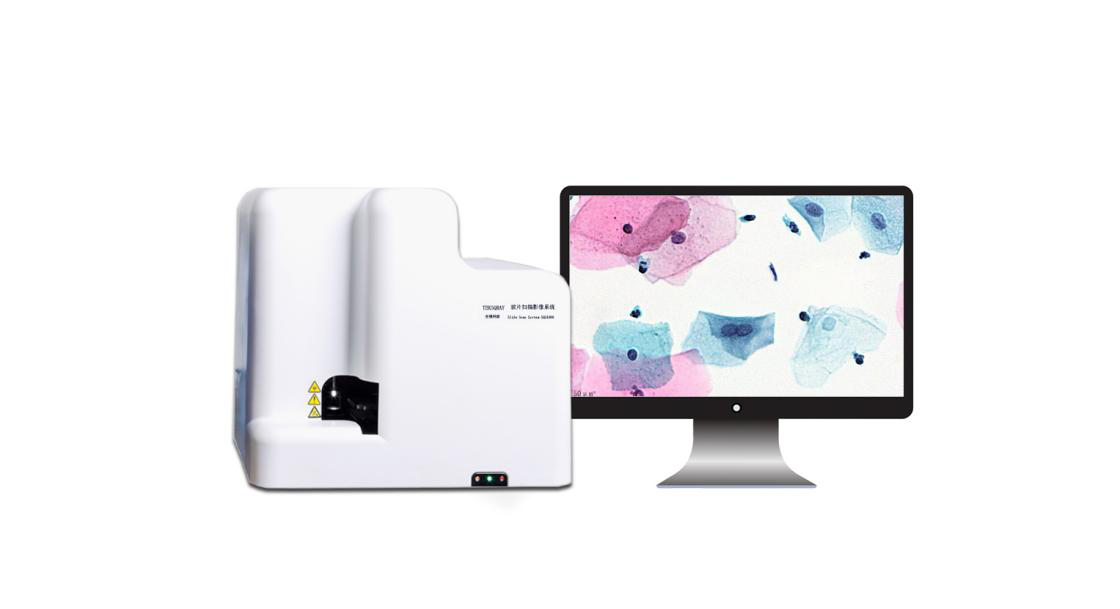
Slide scanning imaging system -20 times/40 times (single slide loading)
The latest self-developed and designed slide scanning image analysis system of Shengqiang Technology has flexible objective lens configuration, high-precision scanning platform, and large-size scanning; it generates a digital slice with a full field of view, which can digitize, complete and permanently store slide information . Experts can browse from a macro image, and then place it in any position at any magnification to observe the slice details.
The latest self-developed and designed slide scanning image analysis system of Shengqiang Technology has flexible objective lens configuration, high-precision scanning platform, and large-size scanning; it generates a digital slice with a full field of view, which can digitize, complete and permanently store slide information . Experts can browse from a macro image, and then place it in any position at any magnification to observe the slice details.
Details
Features
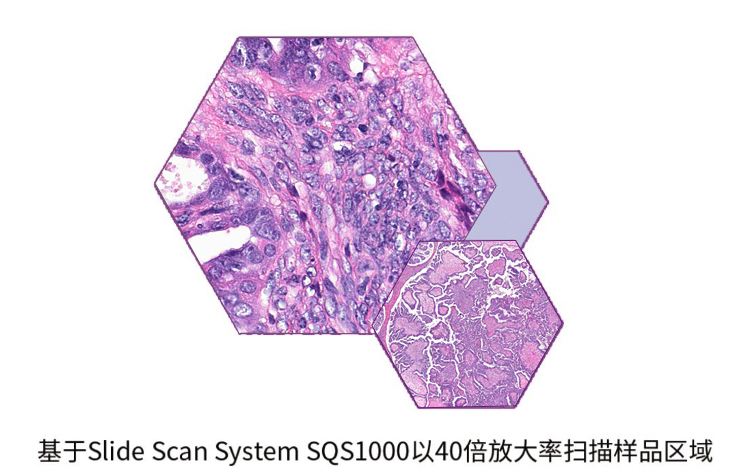
High definition: SlideScan can scan the entire slide quickly and seamlessly. It has high scanning quality and resolution, 20 times is 0.24um/pixel; 40 times is 0.12um/pixel.
Ultra-fast: The slide scanning image analysis system has a unique scanning method. The scanning speed is 30s at 20 times and 85s at 40 times. (15*15mm sample area, higher scanning speed can be customized).
High precision: A closed-loop piezoelectric ceramic motor controlled by nano-displacement controls the Z axis. Focus quickly and accurately.
Scanning control software & Reading software
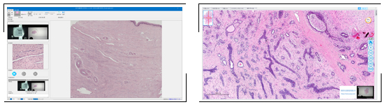
The above system is the supporting software of the slide scanning image analysis system and uses the scanning control software, which can realize the accurate positioning of the slide sample, real-time scanning and imaging point by point, and real-time image stitching and storage. The whole scanning process is automatic, easy to operate, fast and convenient. Use the reading software to open the digital pathology section for viewing and editing. The software supports image formats: sdpc, jpg, bmp, tif, png, rgb888 original image formats, etc. It supports functions such as drawing, adding text, canceling, screenshots, saving information, etc. It supports fixed-point zooming and dragging of the entire image with high response speed.
Main application areas
Pathological slice digitization
Digital information is easier to organize and browse
Can be stored for a long time and save space for storing slides
Pathological diagnosis is easy to read
Remote pathology consultation
Independent of time and space, remote consultation on the Internet
Film reading seminar
Combine with pathology information system to realize true digital pathology information, which can be discussed by multiple people online.
Pathology course teaching
Make tissue slides into digital slices and apply them to histology teaching.
Artificial Intelligence (AI) Pathological Diagnosis
Solve the problem of insufficient cytology doctors
Solve the problem of reading efficiency
Solve the problem of uneven level of reading doctors
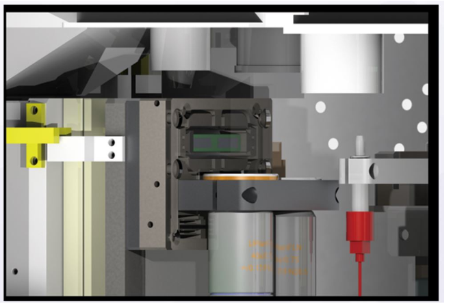
Core technologies
The slide scanning image analysis system has a full-magnification objective lens configuration, which can manually load a single lens, and has a real dual objective lens system. The objective lens is optional, and the Z axis is controlled by an ice ring piezoelectric ceramic motor controlled by nano-displacement. The focusing mechanism is a piezoelectric nano objective lens platform, with a total stroke>150um, a small signal (20um stroke, positioning time <40ms), a resolution of 10nm; fast and precise focus positioning.